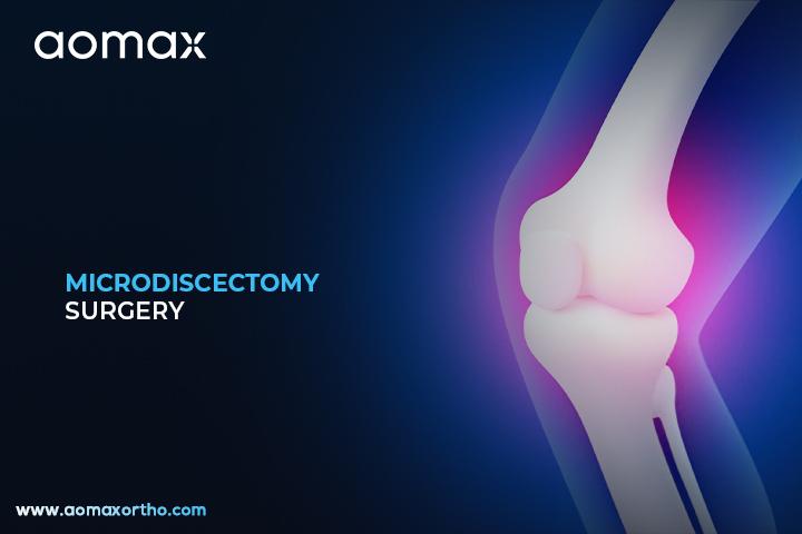
Orthopedic Microdiscectomy – Restoring Spine Function and Reducing Sciatica
Introduction to Microdiscectomy
Microdiscectomy is a minimally invasive surgical procedure to remove herniated disc material pressing on spinal nerves. It is performed to relieve leg or back pain caused by nerve compression. This surgery uses a small incision and specialized tools to minimize tissue damage and speed recovery.
Surgical Process of Microdiscectomy
During microdiscectomy, the surgeon makes a tiny incision near the affected spine area. A surgical microscope or magnifying loupes are used to visualize the disc and nerve roots clearly. Special instruments such as retractors, curettes, and microforceps remove the herniated disc material carefully. Imaging guidance ensures precise removal while avoiding injury to surrounding tissues. The procedure allows for minimal disruption to muscles and ligaments. Patients typically experience faster recovery and less postoperative pain compared to traditional open discectomy.
When is Microdiscectomy Needed?
- To relieve sciatica or radiating leg pain caused by herniated discs.
- To reduce numbness, tingling, or weakness from compressed spinal nerves.
- To improve mobility and daily function when conservative treatments fail.
- To prevent long-term nerve damage from persistent compression.
Products Related to Microdiscectomy
- Kerrison Punch
- MIS Tubular Retractor System
- Spinal Curettes
- Rongeur Forceps
- Hemostatic Bipolar Forceps
- Spinal Bone Punch
- Neurosurgical Punch
- Laminectomy Punch
- Orthopedic Kerrison
- Kerrison Bone Cutter
Worldwide Microdiscectomy Statistics
- United States
- Germany
- Japan
- India
- United Kingdom
Advantages of Microdiscectomy Surgery
- Minimally invasive with smaller incisions and less tissue trauma.
- Rapid pain relief from nerve compression.
- Shorter hospital stay and faster return to daily activities.
- Lower risk of postoperative complications compared to open surgery.
- Preserves spinal stability while targeting the damaged disc precisely.
Aomax Ortho is a globally leading manufacturer of microdiscectomy-related surgical instruments and implants, supporting safe and effective spine surgeries worldwide.
Get Connected:
+91 98989 50530 | exports@aomaxortho.com | www.aomaxortho.com
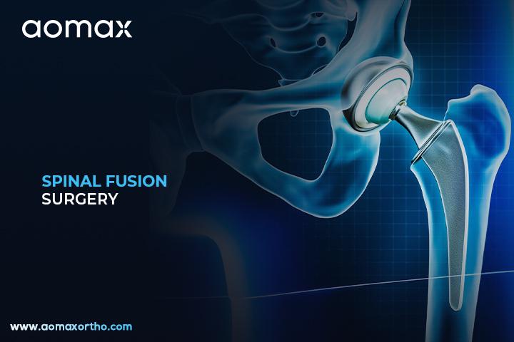
Spinal Fusion – Advanced Orthopedic Surgery for Spine Stability
Spinal Fusion Introduction
Spinal fusion is a surgical procedure that joins two or more vertebrae in the spine. It helps stabilize the spine and reduce pain caused by conditions like scoliosis, degenerative disc disease, or spinal fractures. This surgery limits movement in the affected area to promote proper bone healing and alignment.
Precision Spine Stabilization – Understanding Spinal Fusion Surgery
During spinal fusion, the surgeon removes the damaged disc or prepares the vertebrae to be fused. Bone grafts, either from the patient or donor material, are placed between the vertebrae to facilitate fusion. Metal screws, rods, and plates are used to secure the vertebrae during the healing process. Imaging techniques such as X-ray or fluoroscopy guide the placement of implants accurately. The surgery may be performed using open or minimally invasive techniques depending on the case. Over time, the vertebrae grow together into a solid, stable structure, relieving pain and improving spinal function.
Why it’s done?
- To relieve chronic back or leg pain caused by degenerative disc disease.
- To correct spinal deformities such as scoliosis or kyphosis.
- To stabilize the spine after fractures or spinal injuries.
- To treat conditions like spondylolisthesis or spinal instability.
Products Related to Spinal Fusion
- Spine Endoscope
- Spine Dilator
- SS Spinal Trocar Sleeve
- Orthopedic Hammer
- Spine Sheath
- Spine Grasping Forceps
- Spine Elevator
- Spinal Needle
- Bone Drill Machine
- Arthroscopy Set
Top 5 Countries of Spinal Fusion Surgery
- United States
- Germany
- Japan
- India
- United Kingdom
Spinal Fusion Benefits
- Provides long-term spinal stability and reduces motion at painful segments.
- Corrects spinal deformities and improves posture.
- Relieves nerve compression and associated pain.
- Prevents further spinal degeneration in affected areas.
- Enhances overall quality of life by allowing patients to resume daily activities.
Aomax Ortho is a globally leading manufacturer of spinal fusion-related surgical implants and instruments, trusted by orthopedic surgeons worldwide.
Get Connected:
+91 98989 50530 | exports@aomaxortho.com | www.aomaxortho.com
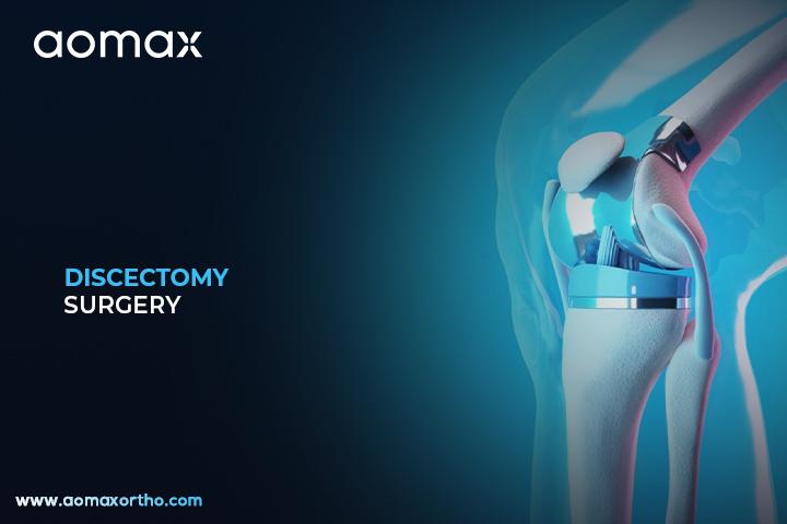
Discectomy – Minimally Invasive Orthopedic Surgery for Spine Health
Overview of Discectomy Surgery
Discectomy is a surgical procedure used to remove part of a damaged or herniated disc in the spine. It helps relieve pressure on nerves that cause pain, weakness, or numbness. This surgery is commonly performed when non-surgical treatments do not provide relief.
Surgical Process of Discectomy
During a discectomy, the surgeon makes a small incision in the back or neck to access the affected spine area. Special instruments such as retractors, surgical microscopes, and curettes are used to carefully remove the damaged disc portion. The procedure may be done as an open surgery or minimally invasive technique depending on the case. By removing the disc material pressing on nerves, this surgery restores mobility and reduces nerve irritation. Surgeons often use imaging guidance to ensure precision throughout the process. Recovery is usually faster when minimally invasive methods are applied.
When is Discectomy Needed?
- To relieve persistent leg or arm pain caused by a herniated disc.
- To reduce numbness, tingling, or weakness from compressed spinal nerves.
- To restore mobility and improve quality of life when conservative treatments fail.
- To prevent long-term nerve damage caused by pressure on the spinal cord or nerve roots.
Essential Tools for Discectomy Surgery
- Spinal Retractors
- Kerrison Punch
- Nerve Root Retractor
- Disc Punch
- Pituitary Forceps
- Curettes
- Surgical Microscope
- Suction Cannula
- High-Speed Drill System
- Bipolar Cautery
Global Presence of Discectomy Surgery
- United States
- Germany
- Japan
- India
- United Kingdom
Discectomy Benefits
- Provides fast and lasting relief from nerve pain.
- Helps restore normal function and mobility of the spine.
- Reduces dependence on long-term pain medications.
- Allows patients to return to daily activities and work more quickly.
- Prevents permanent nerve injury by relieving pressure in time.
Aomax Ortho is a globally leading manufacturer of Discectomy-related surgical instruments, supporting safe and effective spine surgeries worldwide.
Get Connected:
+91 98989 50530 | exports@aomaxortho.com | www.aomaxortho.com
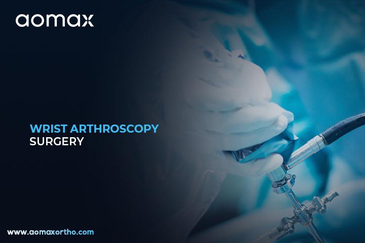
Wrist Arthroscopy – Effective Orthopedic Surgery for Pain & Injury Relief
Understanding Wrist Arthroscopy
Wrist arthroscopy is a minimally invasive surgery that allows doctors to see inside the wrist joint using a small camera. It helps diagnose and treat many wrist problems without making large cuts. This procedure is safer and usually has a quicker recovery time compared to open surgery.
Surgical Technique of Wrist Arthroscopy
During wrist arthroscopy, the surgeon makes tiny cuts around the wrist to insert a small arthroscope and other surgical tools. A camera inside the arthroscope sends clear images of the wrist joint to a monitor, helping the surgeon work with high accuracy. The surgery is often done using specialized tools like arthroscopic shavers, graspers, and probes. Saline solution is used to keep the joint open and provide a clear view. Surgeons can repair ligaments, remove loose bodies, or treat cartilage damage during this procedure. This technique is widely valued for reducing trauma to surrounding tissues and allowing faster rehabilitation.
When is Wrist Arthroscopy Needed?
- To repair torn ligaments inside the wrist joint.
- To treat cartilage damage caused by injury or arthritis.
- To remove loose bone or tissue fragments that cause pain.
- To diagnose unexplained wrist pain not visible on scans.
- To treat wrist instability or stiffness.
Equipment Used in Wrist Arthroscopy
- Arthroscope with HD camera system
- Arthroscopy shaver handpiece & blades
- Arthroscopy probe (hook, palpator)
- Grasper instruments (arthroscopic grasper)
- Arthroscopic scissors
- Trocar and cannula set for wrist portals
- Fluid management pump & tubing
- Light source and fiber‐optic cable
- Retractors and curettes for wrist joint work
- Suture passers and knot pushers
Global Leaders in Wrist Arthroscopy Surgery
- United States
- Germany
- Japan
- United Kingdom
- India
Wrist Arthroscopy Benefits
- Provides a clear view of small wrist structures for accurate diagnosis.
- Minimizes surgical cuts, reducing the risk of infection.
- Shorter recovery period compared to open wrist surgery.
- Less pain and scarring for patients after surgery.
- Helps treat multiple wrist conditions in a single procedure.
- Improves joint function and hand mobility effectively.
Aomax Ortho is a globally leading manufacturer of Wrist Arthroscopy related products, offering advanced solutions to support safe and effective orthopedic surgeries.
Get Connected:
+91 98989 50530 | exports@aomaxortho.com | www.aomaxortho.com
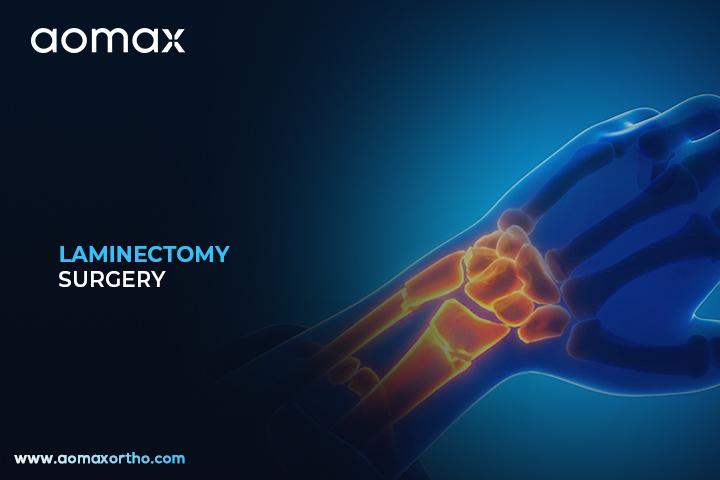
Laminectomy – Minimally Invasive Orthopedic Surgery for Spine Health
Introduction to Laminectomy
Laminectomy is a surgical procedure performed to relieve pressure on the spinal cord or nerves. It involves removing a portion of the vertebra called the lamina. This surgery is often recommended for patients with spinal stenosis or other spinal disorders.
Surgical Process and Role of Laminectomy
During a laminectomy, the surgeon makes an incision over the affected spine area and carefully moves aside muscles to access the vertebra. Specialized surgical instruments are used to remove the lamina, which reduces compression on nerves. Sometimes, bone spurs or thickened ligaments are also removed to create more space in the spinal canal. The procedure may be performed on the cervical, thoracic, or lumbar spine depending on the condition. Advanced orthopedic tools and implants may be required if spinal stabilization is necessary. This surgery helps restore mobility, reduces pain, and improves the patient’s quality of life.
When Surgery Is Needed
- To relieve nerve compression caused by spinal stenosis
- To remove bone spurs or thickened tissues pressing on the spinal cord
- To treat herniated or ruptured discs causing severe symptoms
- To improve mobility and reduce chronic back or leg pain
- To address spinal tumors or abnormal growths affecting the spine
Essential Surgical Instruments in Laminectomy
- High-quality retractors (muscle retractors / nerve root retractors) — necessary to expose the lamina
- Bone cutting instruments (e.g. osteotomes, spinal curettes)
- High-speed drill systems (for laminectomy window cutting)
- Suction / irrigation sets to keep the surgical field clean
- Bone holding forceps
- Rongeurs — e.g. Kerrison Rongeurs — to remove lamina or bone spurs
- Elevators (spinal elevators) to help mobilize soft tissue
- Hemostatic tools / bipolar cautery to control bleeding
- Spine-specific curettes or scrapers for ligament removal
- Spinal implant systems (if stabilization or fusion is needed after laminectomy)
Countries Leading in Laminectomy
- United States
- Germany
- Japan
- India
- Brazil
Clinical Outcomes of Laminectomy
- Provides long-term relief from nerve compression symptoms
- Improves overall spinal stability when combined with fusion techniques
- Restores the ability to walk, sit, or stand without severe pain
- Reduces dependence on pain medications for chronic spinal issues
- Enhances quality of life and daily functional ability
- Allows surgical correction of serious spinal abnormalities or tumors
Aomax Ortho is a globally leading manufacturer of laminectomy-related products, offering high-quality surgical instruments and solutions to support advanced spinal procedures.
Get Connected:
+91 98989 50530 | exports@aomaxortho.com | www.aomaxortho.com
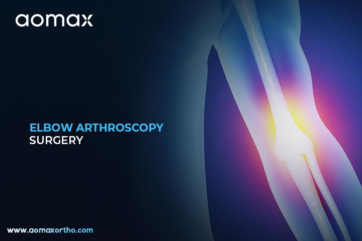
Elbow Arthroscopy – Effective Orthopedic Surgery for Pain & Injury Repair
Overview of Elbow Arthroscopy
Elbow arthroscopy is a minimally invasive surgical procedure used to diagnose and treat problems inside the elbow joint. Surgeons make small cuts in the skin and insert a tiny camera to see inside the joint. This technique allows precise treatment with less pain and faster recovery compared to open surgery.
Inside the Elbow Arthroscopy Procedure
During elbow arthroscopy, surgeons create small portals around the elbow to insert the arthroscope and surgical tools. A camera attached to the arthroscope projects a clear view of the joint on a monitor, helping the surgeon identify damaged tissue, loose bodies, or inflammation. Saline solution is used to expand the joint, giving better visibility during the procedure. Specialized instruments such as shavers, graspers, and radiofrequency devices are used to trim or remove damaged tissue. Surgeons can also repair ligaments or smooth rough bone surfaces through the same small incisions. The method ensures less trauma to surrounding tissues and quicker functional recovery.
Medical Reasons for Elbow Arthroscopy
- To remove loose bone or cartilage fragments inside the elbow joint
- To treat stiffness or limited motion caused by scar tissue
- To repair damaged ligaments or tendons
- To treat arthritis-related problems like bone spurs
- To manage chronic elbow pain that does not improve with non-surgical treatments
Surgical Tools Used in Elbow Arthroscopy
- Arthroscope – 4 mm
- Arthroscopy Sheath
- Arthroscopy Shaver System
- Arthroscopy RF Cautery Machine
- Orthopedic Arthro Pump
- Digital Tourniquet
- Orthopedic Dilator
- Orthopedic Hook
- Arthroscopy Probe
- Arthroscopy Cannula
Countries Performing the Most Elbow Arthroscopies
- United States
- Germany
- Japan
- United Kingdom
- India
Advantages of Elbow Arthroscopy
- Provides accurate diagnosis and treatment with minimal tissue damage
- Results in faster recovery and earlier return to daily activities
- Reduces post-operative pain compared to open surgery
- Lowers the risk of complications like infection and stiffness
- Improves joint movement and overall elbow function
Aomax Ortho is a globally trusted manufacturer of Elbow Arthroscopy-related products, offering advanced solutions to support surgeons in achieving successful outcomes.
Get Connected:
+91 98989 50530 | exports@aomaxortho.com | www.aomaxortho.com
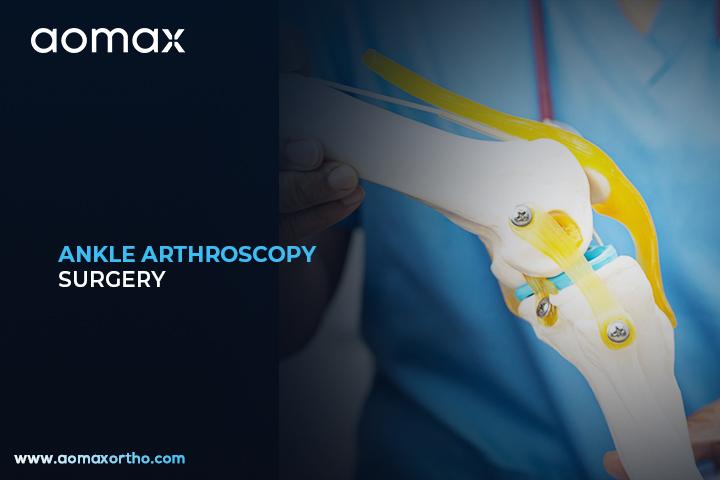
Ankle Arthroscopy – Trusted Orthopedic Surgery for Sports & Trauma Care
Overview of Ankle Joint Surgery
Ankle arthroscopy is a minimally invasive surgery used to treat problems inside the ankle joint. Surgeons use a small camera and special instruments inserted through tiny cuts to see and repair the joint. This surgery helps reduce pain, improve movement, and speed up recovery.
How Ankle Arthroscopy is Performed
During ankle arthroscopy, a surgeon makes small incisions around the ankle to insert an arthroscope and thin surgical tools. The arthroscope is a tiny camera that provides a clear view inside the joint on a monitor. Surgeons can then remove damaged tissue, smooth rough cartilage, or repair ligaments. Saline fluid is often used to expand the joint for better visibility. Advanced instruments like shavers and radiofrequency devices allow precise treatment of affected areas. This technique is less invasive than open surgery and usually results in faster healing and fewer complications.
Common Health Issues Requiring This Surgery
- To treat chronic ankle pain caused by cartilage damage.
- To remove loose bone or cartilage fragments inside the joint.
- To repair torn or weakened ligaments.
- To treat ankle impingement, which restricts joint movement.
- To manage joint inflammation or infection not improving with other treatments.
Surgical Tools Used in Ankle Arthroscopy
- Arthroscope – 4 mm
- Arthroscopy Sheath
- Arthroscopy Trocar
- Arthroscopy Punch
- Arthroscopy Grasper
- Arthroscopy Scissors
- Arthroscopy Shaver System
- Arthroscopy RF Cautery Machine
- Fluid Management System (Arthro Pump)
- Cannula System
Countries with the Highest Procedure Rates
- United States
- Germany
- Japan
- United Kingdom
- India
Clinical Benefits of Ankle Arthroscopy
- Minimally invasive surgery with smaller cuts and less scarring.
- Faster recovery compared to traditional open ankle surgery.
- Reduced risk of infection due to smaller incisions.
- Precise visualization of the joint, allowing accurate treatment.
- Helps restore ankle strength, stability, and long-term mobility.
Aomax Ortho is a globally leading manufacturer of Ankle Arthroscopy related products, offering advanced and reliable solutions for orthopedic surgeries worldwide.
Get Connected:
+91 98989 50530 | exports@aomaxortho.com | www.aomaxortho.com
Hip Arthroscopy – Advanced Orthopedic Surgery for Hip Joint Preservation
Understanding Hip Arthroscopy
Hip arthroscopy is a minimally invasive surgery used to treat problems inside the hip joint. Surgeons use a tiny camera and special instruments inserted through small cuts to see and repair the hip. This procedure helps patients recover faster with less pain compared to open surgery.
Advanced Hip Arthroscopy Technique for Joint Preservation
During hip arthroscopy, surgeons insert an arthroscope through a small incision to clearly view the hip joint. Specialized instruments are then used to trim damaged cartilage, remove loose fragments, or repair torn labrum tissue. The procedure may also include smoothing irregular bone surfaces to improve joint movement. Continuous fluid irrigation is used to expand the joint and provide a clear view for precise work. High-definition visualization systems guide surgeons to perform accurate and controlled repairs. This technique helps preserve natural hip structure and reduces the need for more invasive surgeries in the future.
Conditions Treated with Hip Arthroscopy
- To repair a torn hip labrum and restore joint stability.
- To remove loose bodies of cartilage or bone causing pain.
- To treat femoroacetabular impingement (FAI) and prevent further damage.
- To address early stages of arthritis and delay joint replacement.
Products Related to Hip Arthroscopy
- Arthroscope
- Arthroscopy Cannula
- Arthroscopy Trocar
- Arthroscopy Hook Scissors
- Arthroscopy Punch
- Arthroscopy Shaver System
- Arthroscopy RF Cautery System
- Arthroscopy Probe
- Fiber Optic Light Cable
- Arthro Pump
Worldwide Adoption of Hip Arthroscopy
- United States
- Germany
- United Kingdom
- Japan
- India
How Patients Benefit from Hip Arthroscopy
- Minimally invasive approach with faster recovery time.
- Reduced risk of infection compared to open surgery.
- Preservation of natural joint function and structure.
- Improved accuracy due to enhanced visualization of the hip joint.
- Effective pain relief and improved mobility in daily activities.
Aomax Ortho is a leading manufacturer of Hip Arthroscopy related products, supporting surgeons worldwide with high-quality and reliable instruments.
Get Connected:
+91 98989 50530 | exports@aomaxortho.com | www.aomaxortho.com
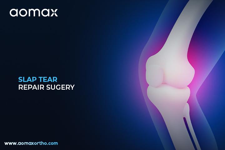
Orthopedic SLAP Tear Repair – Shoulder Joint Stability & Function Restoration
Overview of SLAP Tear Repair
SLAP tear repair is a surgery used to fix a tear in the labrum of the shoulder joint. The labrum is a ring of cartilage that helps stabilize the shoulder. This surgery is commonly done when the tear causes pain, weakness, or difficulty in shoulder movement.
Advanced Insights into SLAP Tear Repair
During SLAP tear repair, the surgeon uses an arthroscope, a small camera inserted through tiny incisions, to see inside the shoulder. Specialized instruments are used to reattach the torn labrum to the bone using sutures or anchors. The procedure is usually performed under regional or general anesthesia. Surgeons may use absorbable or non-absorbable suture anchors depending on the tear type. The minimally invasive approach helps reduce tissue damage and recovery time. This surgery improves shoulder strength, stability, and range of motion for patients with labral injuries.
When is SLAP Tear Repair Needed?
- To treat shoulder instability caused by a torn labrum.
- To relieve persistent shoulder pain that does not improve with physiotherapy or medications.
- To restore strength and motion for athletes with repetitive overhead movements.
- To repair labral damage from trauma, dislocation, or heavy lifting injuries.
Products Related to SLAP Tear Repair
- Arthroscope
- Arthroscopy Pump
- Arthroscopy RF Cautery System
- Knotless Suture Anchor
- PEEK Suture Anchor with Fiber
- All-Suture Anchor
- Fiber Wire
- Fiber Tape
- Arthroscopy Cannula
- Arthroscopy Hook Scissors
Countries Performing the Most SLAP Tear Repair
- United States
- Germany
- United Kingdom
- Japan
- India
Key Outcomes of SLAP Tear Repair
- Provides long-term stability to the shoulder joint.
- Improves overhead motion and athletic performance.
- Reduces the risk of recurrent shoulder dislocation or subluxation.
- Helps in faster recovery compared to open surgical techniques.
- Decreases the chance of future shoulder arthritis caused by untreated labral tears.
Aomax Ortho is a leading manufacturer of SLAP Tear Repair-related products, offering advanced arthroscopy instruments and implants to support successful orthopedic surgeries.
Get Connected:
+91 98989 50530 | exports@aomaxortho.com | www.aomaxortho.com

Bankart Repair – Effective Surgical Solution for Shoulder Instability
Overview of Bankart Repair Surgery
Bankart Repair is a surgical procedure aimed at stabilizing the shoulder joint by repairing a torn labrum. This tear often results from recurrent shoulder dislocations. The surgery restores the shoulder’s normal anatomy, reducing the risk of future dislocations.
Restoring Shoulder Stability – The Bankart Repair Procedure
Bankart Repair is typically performed using minimally invasive arthroscopic techniques. Small incisions are made around the shoulder to insert a camera and specialized instruments. The torn labrum is reattached to the glenoid (shoulder socket) using suture anchors. This reattachment restores the bumper effect of the labrum, keeping the humeral head centered during shoulder motion. The procedure aims to stabilize the shoulder joint and prevent future dislocations. Post-surgery, a structured rehabilitation program is essential for optimal recovery.
When is Bankart Repair Needed?
- To repair a torn labrum resulting from recurrent shoulder dislocations.
- To stabilize the shoulder joint and prevent future dislocations.
- To alleviate pain associated with shoulder instability.
- To restore normal shoulder function and range of motion.
Key Products for Bankart Repair Surgery
- Arthroscopic Suture Anchors
- Suture Passing Instruments
- Arthroscopic Shavers and Debridement Tools
- Shoulder Positioners
- Cannulas for Arthroscopy
- Arthroscopic Camera Systems
- Knot Pushers
- Bioabsorbable Anchors
- RF Ablation Probes
- Shoulder Portal Instruments
Countries with Highest Bankart Repair Rates
- United States
- Germany
- Japan
- United Kingdom
- Australia
Positive Outcomes of Bankart Repair
- Restores shoulder stability, reducing the risk of future dislocations.
- Alleviates pain associated with shoulder instability.
- Improves range of motion and shoulder function.
- Minimally invasive approach leads to quicker recovery times.
- Allows patients to return to sports and daily activities safely.
Aomax Ortho is a leading manufacturer of Bankart Repair related products, offering reliable and advanced surgical tools for orthopedic procedures.
Get Connected:
+91 98989 50530 | exports@aomaxortho.com | www.aomaxortho.com