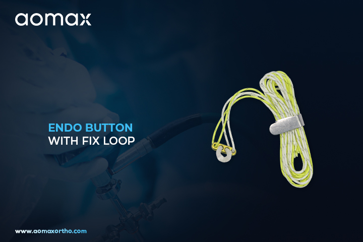
Applications of Endo Button with Fixed Loop in Orthopedic Surgery
Endo Button with Fix Loop:
Endo Button with Fix Loop is an orthopedic fixation implant designed to secure tendon and ligament grafts during reconstructive procedures. Its fixed loop provides a predetermined loop length for consistent graft positioning and cortical fixation. The Endo Button supports stable graft fixation in ligament reconstruction procedures, particularly arthroscopic applications.
Endo Button with Fix Loop History:
Endo Button fixation systems were developed to provide cortical fixation for tendon and ligament grafts during reconstructive orthopedic surgery. Fixed-loop designs were developed to offer a consistent loop length and straightforward graft fixation technique. These fixation systems have become widely used in ligament reconstruction procedures, including ACL reconstruction and sports medicine applications.
Short Description:
Aomax Ortho Endo Button with Fix Loop is designed for cortical fixation of tendon and ligament grafts during orthopedic reconstruction. Aomax Ortho provides this fixation system with a fixed loop configuration that maintains a predetermined loop length during graft placement. The button supports cortical fixation while the fixed loop helps maintain consistent graft positioning. Its compact design is suitable for arthroscopic ligament reconstruction procedures. The device can be selected for ACL and other reconstructive procedures where fixed-loop graft fixation is required.
Surgeries Related to Product:
- ACL reconstruction
- PCL reconstruction
- Knee ligament reconstruction
- Arthroscopic ligament reconstruction
- Tendon graft fixation
- Sports injury reconstruction
Instructions for Use:
- Select the appropriate Endo Button with Fix Loop according to the graft and surgical technique.
- Prepare the graft and bone tunnel according to the established surgical procedure.
- Pass the fixation system through the prepared tunnel as directed by the applicable technique.
- Confirm proper cortical engagement and button positioning.
- Position and tension the graft according to the surgical technique.
- Verify graft placement and fixation before completing the reconstruction.
- Use only by qualified healthcare professionals according to the applicable instructions for use.
Top 10 Countries of Surgeries:
- United States
- Germany
- United Kingdom
- India
- France
- Japan
- Italy
- South Korea
- Australia
- Canada
Clinical Benefits:
- Provides cortical fixation for tendon and ligament grafts.
- Fixed loop offers a predetermined loop configuration.
- Supports consistent graft positioning during reconstruction.
- Compact design supports use in arthroscopic procedures.
- Helps stabilize the graft during ligament reconstruction.
- Suitable for ACL and other ligament reconstruction applications.
- Provides a practical graft fixation option for sports medicine procedures.
Market Demand and Global Trends:
Fixed-loop graft fixation systems continue to be used in orthopedic and sports medicine procedures requiring cortical fixation. The established configuration provides a predetermined loop length for graft fixation and is suitable for surgical techniques where a fixed loop is preferred. Increasing use of arthroscopic ligament reconstruction contributes to demand for specialized graft fixation implants. ACL reconstruction and other sports-related ligament procedures remain important applications for Endo Button fixation systems.
Aomax Ortho Endo Button with Fix Loop Description:
Aomax Ortho is a leading manufacturer and supplier of Endo Button with Fix Loop, providing orthopedic graft fixation systems designed for cortical fixation and stable tendon or ligament graft positioning during reconstructive procedures. The system is suitable for arthroscopic, orthopedic, and sports medicine applications requiring fixed-loop graft fixation.
Get Connected:
+91 98989 50530 | exports@aomaxortho.com | www.aomaxortho.com

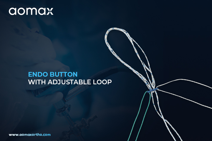
Understanding Adjustable Loop Endo Button in ACL Reconstruction
Introduction to Endo Button with Adjustable Loop:
Endo Button with Adjustable Loop is an orthopedic fixation implant designed to secure tendon and ligament grafts during reconstructive procedures. Its adjustable loop allows graft fixation and tensioning according to the requirements of the surgical technique. The Endo Button provides cortical fixation while the adjustable loop supports controlled graft positioning.
Endo Button with Adjustable Loop History:
Endo Button fixation systems were developed to provide cortical fixation for tendon and ligament grafts during reconstructive orthopedic surgery. Adjustable loop technology was introduced to provide greater flexibility in graft positioning and tension adjustment compared with fixed-loop configurations. These systems are now commonly used in arthroscopic ligament reconstruction procedures, particularly ACL reconstruction.
Short Description:
Aomax Ortho Endo Button with Adjustable Loop is designed for cortical fixation and controlled positioning of tendon or ligament grafts during reconstruction. Aomax Ortho provides this fixation system with an adjustable loop that allows the surgeon to modify graft length and tension according to the surgical technique. The button supports cortical fixation while the adjustable loop provides flexibility during graft placement. Its configuration is suitable for arthroscopic procedures involving ligament reconstruction and graft stabilization. The system can be selected for procedures where adjustable graft fixation is required.
Surgeries Related to Product:
- ACL reconstruction
- PCL reconstruction
- Knee ligament reconstruction
- Arthroscopic ligament reconstruction
- Tendon graft fixation
- Sports injury reconstruction
Instructions for Use:
- Select the appropriate fixation system according to the graft and surgical technique.
- Prepare the graft and bone tunnel following the established surgical procedure.
- Pass the fixation system through the prepared tunnel as directed by the applicable technique.
- Confirm proper cortical engagement of the Endo Button.
- Adjust the loop according to the required graft positioning and tension.
- Verify graft placement and fixation before completing the reconstruction.
- Use only by qualified healthcare professionals according to the applicable instructions for use.
Top 10 Countries of Surgeries:
- United States
- Germany
- United Kingdom
- India
- France
- Japan
- Italy
- South Korea
- Australia
- Canada
Clinical Benefits:
- Provides cortical fixation for tendon and ligament grafts.
- Adjustable loop allows flexibility in graft positioning.
- Supports controlled graft tensioning during reconstruction.
- Compact configuration is suitable for arthroscopic procedures.
- Supports graft stabilization during ligament reconstruction.
- Titanium or other specified button materials can provide durable fixation according to product configuration.
- Suitable for ACL and other ligament reconstruction applications.
Product Also Known As:
Endo Button with Adjustable Loop, adjustable loop Endo Button, adjustable Endobutton, ACL Endo Button with adjustable loop, adjustable loop graft fixation button, ligament fixation button with adjustable loop, adjustable cortical fixation button, orthopedic Endo Button implant
Frequently Used Product Combination:
- ACL graft
- Femoral fixation system
- Tibial fixation screw
- Titanium interference screw
- PEEK interference screw
- Arthroscopic instruments
- Ligament reconstruction instruments
- Graft preparation instruments
Aomax Ortho Endo Button with Adjustable Loop Description:
Aomax Ortho is a leading manufacturer and supplier of Endo Button with Adjustable Loop, providing orthopedic graft fixation systems designed for cortical fixation and adjustable tendon or ligament graft positioning during reconstructive procedures. The system is suitable for arthroscopic, orthopedic, and sports medicine applications.
Get Connected:
+91 98989 50530 | exports@aomaxortho.com | www.aomaxortho.com
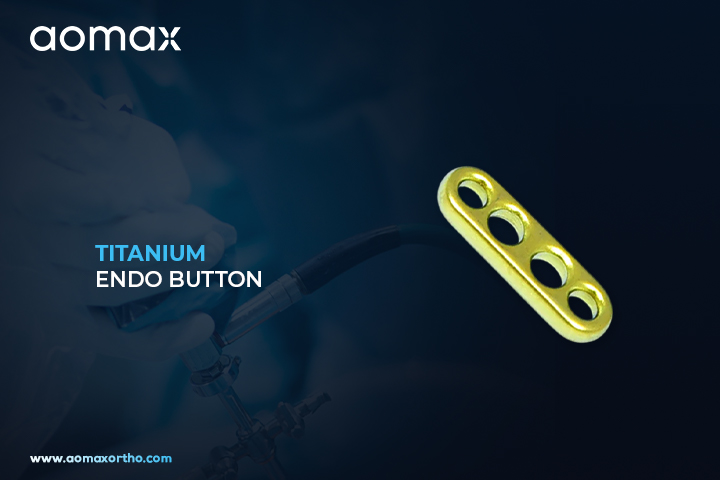
Titanium Endo Button for Ligament Graft Fixation in Orthopedic Surgery
Introduction to Titanium Endo Button:
Titanium Endo Button is an orthopedic fixation implant designed to secure tendon and ligament grafts during reconstructive procedures. It provides cortical fixation to help maintain graft positioning during ligament reconstruction. The titanium construction offers strength, durability, and dependable fixation support for orthopedic applications.
Titanium Endo Button History:
Titanium Endo Button fixation systems were developed to provide cortical fixation for tendon and ligament grafts in reconstructive orthopedic surgery. Titanium has been widely used in orthopedic implants because of its strength, durability, and established biocompatibility. Titanium Endo Buttons are commonly used as part of graft fixation techniques in ligament reconstruction procedures.
Role of Cortical Fixation in Ligament Reconstruction:
Aomax Ortho Titanium Endo Button is designed for cortical fixation of tendon and ligament grafts during orthopedic reconstruction. Aomax Ortho manufactures this titanium fixation implant with a durable structure suitable for ligament stabilization procedures. Its compact button configuration supports secure positioning on the cortical surface during graft fixation. The implant can be used in arthroscopic and reconstructive orthopedic procedures according to the selected surgical technique. It is commonly associated with ACL and other ligament reconstruction applications where reliable graft fixation is required.
Surgeries Related to Product:
- ACL reconstruction
- PCL reconstruction
- Knee ligament reconstruction
- Arthroscopic ligament reconstruction
- Tendon graft fixation
- Sports injury reconstruction
Instructions for Use:
- Select the appropriate implant based on the graft, tunnel dimensions, and surgical technique.
- Prepare the graft and bone tunnel according to the established surgical procedure.
- Pass the fixation system through the prepared tunnel as directed by the applicable technique.
- Confirm proper cortical engagement and Titanium Endo Button positioning.
- Apply appropriate graft tension according to the surgical procedure.
- Verify graft position and fixation before completing the reconstruction.
- Use only by qualified healthcare professionals according to the applicable instructions for use.
Clinical Benefits of Titanium Graft Fixation:
- Provides cortical fixation for tendon and ligament grafts.
- Titanium construction supports mechanical strength and durability.
- Compact configuration supports efficient graft fixation.
- Helps maintain graft positioning during reconstruction.
- Suitable for arthroscopic orthopedic procedures.
- Supports ACL and other ligament reconstruction applications.
- Provides a practical fixation option for sports medicine procedures.
Product Also Known As:
Titanium Endo Button, titanium Endobutton, titanium endo button implant, titanium graft fixation button, titanium cortical fixation button, ACL titanium fixation button, titanium ligament fixation button, orthopedic titanium Endo Button
Market Demand and Global Trends:
Titanium graft fixation implants continue to be used in orthopedic and sports medicine procedures requiring cortical fixation. Growing adoption of arthroscopic and minimally invasive ligament reconstruction supports demand for specialized graft fixation devices. ACL reconstruction remains an important application for Titanium Endo Button fixation systems, particularly in sports medicine. Manufacturers are also developing different fixation configurations to accommodate varying graft types and surgical techniques.
Aomax Ortho Titanium Endo Button Description:
Aomax Ortho is a leading manufacturer and supplier of Titanium Endo Button, providing titanium orthopedic fixation implants designed to support tendon and ligament graft stabilization during reconstructive procedures. The implant is suitable for orthopedic, arthroscopic, and sports medicine applications where cortical graft fixation is required.
Get Connected:
+91 98989 50530 | exports@aomaxortho.com | www.aomaxortho.com
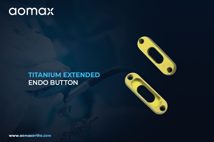
Titanium Extended Endo Button for Reliable Ligament Graft Fixation
Titanium Extended Endo Button
Titanium Extended Endo Button is an orthopedic fixation implant designed for securing tendon or ligament grafts during reconstructive procedures. It provides cortical fixation and supports stable graft positioning during ligament reconstruction. The titanium construction offers strength, durability, and reliable mechanical support for orthopedic applications.
Titanium Extended Endo Button History:
Endo Button fixation technology was developed to provide cortical fixation for tendon and ligament grafts in reconstructive orthopedic surgery. Titanium became a commonly used material for fixation implants because of its strength, durability, and established use in orthopedic applications. Extended Endo Button designs provide additional fixation options for procedures requiring an extended button configuration.
Short Description:
Aomax Ortho Titanium Extended Endo Button is designed to provide secure cortical fixation for tendon and ligament grafts during orthopedic reconstruction. Aomax Ortho offers this titanium fixation implant with a durable construction suitable for orthopedic applications. Its extended configuration provides versatility for graft fixation according to the surgical technique and tunnel requirements. The compact design supports efficient placement during arthroscopic and open reconstructive procedures. It is particularly suitable for ligament reconstruction procedures such as ACL surgery where reliable graft fixation is required.
Surgeries Related to Product:
- ACL reconstruction
- PCL reconstruction
- Knee ligament reconstruction
- Arthroscopic ligament reconstruction
- Tendon graft fixation
- Sports injury reconstruction
Instructions for Use:
- Select the appropriate implant according to the surgical technique and graft requirements.
- Prepare the graft and bone tunnel according to the established surgical procedure.
- Position the fixation device through the prepared tunnel as directed by the surgical technique.
- Confirm appropriate cortical engagement and button positioning.
- Apply suitable graft tension according to the surgeon’s procedure.
- Verify graft position and fixation before completing the surgery.
- Use only by qualified healthcare professionals according to the applicable instructions for use.
Top 10 Countries of Surgeries:
- United States
- Germany
- United Kingdom
- India
- France
- Japan
- Italy
- South Korea
- Australia
- Canada
Clinical Benefits:
- Provides cortical fixation for tendon and ligament grafts.
- Titanium construction supports strength and durability.
- Extended configuration provides versatile graft fixation options.
- Supports stable graft positioning during reconstruction.
- Suitable for arthroscopic orthopedic procedures.
- Compact design supports efficient surgical placement.
- Useful for ACL and other ligament reconstruction applications.
Product Also Known As:
titanium extended endo button, extended endobutton titanium, titanium graft fixation button, extended cortical fixation button
Market Demand and Global Trends:
Titanium fixation implants remain widely used in orthopedic and sports medicine procedures requiring durable graft fixation. Increasing adoption of arthroscopic and minimally invasive ligament reconstruction is supporting demand for specialized fixation devices. ACL reconstruction is an important application area due to the prevalence of sports-related and ligament injuries. Extended Endo Button designs provide additional fixation options for surgeons working with different graft and bone tunnel configurations.
Aomax Ortho Titanium Extended Endo Button Description:
Aomax Ortho is a leading manufacturer and supplier of Titanium Extended Endo Button, offering titanium orthopedic fixation implants designed for tendon and ligament graft stabilization during reconstructive procedures. The device is suitable for orthopedic and sports medicine applications where dependable cortical fixation is required.
Get Connected:
+91 98989 50530 | exports@aomaxortho.com | www.aomaxortho.com
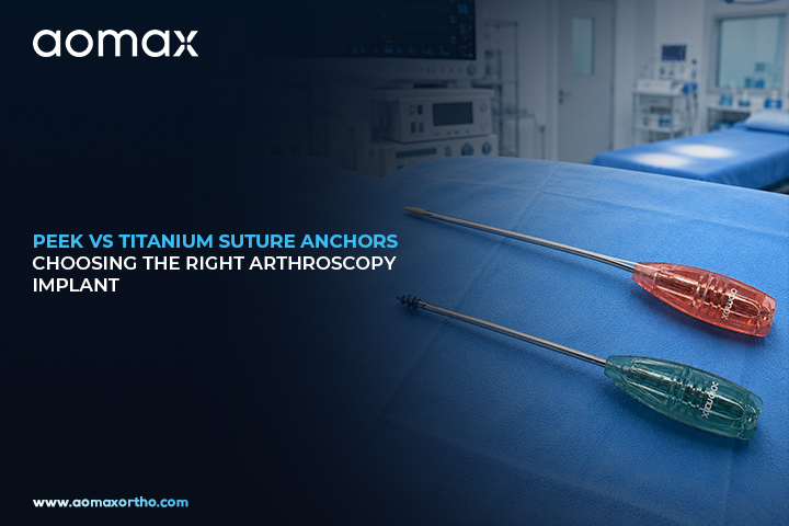
PEEK vs Titanium Suture Anchors – Choosing the Right Arthroscopy Implant
In modern arthroscopic surgery, selecting the right implant material is just as important as surgical technique. Among the most widely used options, PEEK (Polyether Ether Ketone) and Titanium suture anchors stand out for their performance, reliability, and clinical acceptance.
Both materials are extensively used for soft tissue to bone fixation, but they differ significantly in terms of biomechanics, imaging compatibility, and long-term outcomes. Understanding these differences helps surgeons make the right choice based on patient needs and surgical goals.
What Are Suture Anchors?
Suture anchors are orthopedic implants used to attach soft tissues like tendons and ligaments to bone. They are commonly used in:
- Rotator cuff repair
- ACL and PCL reconstruction
- Labral repair (shoulder and hip)
- Tendon reattachment procedures
The choice of anchor material can directly impact healing, imaging, and revision surgery.
What is PEEK?
PEEK (Polyether Ether Ketone) is a high-performance medical-grade polymer widely used in orthopedic implants.
Key Advantages of PEEK Anchors:
- Radiolucent – Does not interfere with X-rays, MRI, or CT scans
- Biocompatible – Minimal inflammatory response
- Elastic modulus similar to bone – Reduces stress shielding
- No corrosion risk – Long-term stability
PEEK anchors are especially preferred in cases where post-operative imaging and long-term monitoring are critical.
What is Titanium?
Titanium is a strong, lightweight metal that has been used in orthopedic implants for decades.
Key Advantages of Titanium Anchors:
- High mechanical strength – Excellent pull-out resistance
- Proven clinical history – Widely trusted material
- Osseointegration – Strong bonding with bone
- Durability – Ideal for high-load applications
Titanium anchors are often used in demanding cases where maximum strength is required.
PEEK vs Titanium – Key Differences
| Feature | PEEK Anchors | Titanium Anchors |
|---|---|---|
| Imaging | Radiolucent (clear imaging) | Can cause imaging artifacts |
| Strength | High but slightly flexible | Very high rigidity |
| Bone Interaction | No osseointegration | Strong bone integration |
| Revision Surgery | Easier to manage | Can be difficult to remove |
| Corrosion | No corrosion | Extremely resistant but metallic |
| Elasticity | Similar to bone | Much stiffer than bone |
Which One Should You Choose?
The choice between PEEK and Titanium suture anchors depends on several clinical factors:
- Choose PEEK when:
- Post-op imaging clarity is important
- Reduced stress shielding is desired
- Revision surgery may be required
- Choose Titanium when:
- Maximum strength and fixation are needed
- High-load or complex repairs are involved
- Long-term bone integration is preferred
Aomax Approach to Implant Innovation
At Aomax, we understand that no single material fits every surgical scenario. That’s why we offer both PEEK and Titanium suture anchor systems, ensuring surgeons have the flexibility to choose the right implant for each procedure.
Our implants are designed with:
- Precision engineering for strong fixation
- Surgeon-friendly insertion systems
- Consistent quality and performance
- Compatibility across multiple arthroscopic procedures
Conclusion
Both PEEK and Titanium suture anchors play a vital role in modern arthroscopy. While PEEK offers imaging advantages and biomechanical compatibility, Titanium provides unmatched strength and long-term stability.
Choosing the right anchor ultimately depends on surgical goals, patient anatomy, and procedural demands. With advanced solutions from Aomax, surgeons can confidently select the ideal implant for optimal outcomes.
Get Connected:
+91 98989 50530 | ortho@advinhealthcare.com | www.advinortho.com
High-Strength Fiber Sutures Manufacturer in India for Orthopedic & Arthroscopy Surgeries
Fiber sutures are a critical component in modern orthopedic and arthroscopic procedures, enabling strong, reliable soft tissue fixation across a wide range of surgical applications. From ligament reconstruction to rotator cuff repair, high-strength sutures play a key role in ensuring stability, durability, and optimal healing.
As a trusted high-strength fiber sutures manufacturer in India, Aomax provides advanced suture solutions designed to meet the demanding requirements of orthopedic and sports medicine surgeries.
What are Fiber Sutures?
Fiber sutures are specialized surgical sutures made from ultra-high molecular weight polyethylene (UHMWPE) and other high-performance materials. These sutures are engineered to provide superior tensile strength, flexibility, and resistance to breakage.
Unlike conventional sutures, fiber sutures are designed specifically for high-load orthopedic applications where durability and secure fixation are essential.
Key Applications in Orthopedic & Arthroscopy Surgeries
Fiber sutures are widely used across multiple procedures, including:
- ACL and PCL reconstruction
- Rotator cuff repair
- Labral repair (shoulder and hip)
- Meniscus repair
- Tendon and ligament fixation
Their versatility makes them an essential component in both open and minimally invasive surgical techniques.
Importance of High-Strength Sutures in Surgery
In orthopedic procedures, sutures must withstand significant mechanical stress during and after surgery.
Key Requirements:
- High tensile strength to prevent breakage
- Excellent knot security
- Minimal elongation under load
- Smooth handling for surgeons
- Biocompatibility for safe implantation
High-strength fiber sutures meet these requirements, ensuring reliable performance even in demanding conditions.
Advantages of Fiber Sutures
Modern fiber sutures offer several advantages over traditional materials:
- Superior strength and durability
- Enhanced knot security
- Resistance to fraying and abrasion
- Improved handling characteristics
- Compatibility with suture anchors and fixation devices
These benefits contribute to better surgical outcomes and reduced risk of repair failure.
Role in Arthroscopy Procedures
In arthroscopic surgeries, fiber sutures are commonly used with anchors and fixation systems.
Key Uses:
- Securing soft tissue to bone
- Creating stable fixation constructs
- Supporting multi-anchor repair techniques
- Enabling knotless fixation systems
Their high performance is essential in minimally invasive procedures where precision and reliability are critical.
Aomax Fiber Suture Solutions
Aomax offers a range of high-strength fiber sutures designed for orthopedic and arthroscopy applications.
Product Features:
- Ultra-high tensile strength
- Excellent knot holding capability
- Smooth and flexible structure
- Biocompatible materials
- Consistent performance
These sutures are engineered to meet the needs of modern surgical techniques and high-demand procedures.
Manufacturing Quality & Standards
As a reliable manufacturer, Aomax ensures:
- Strict quality control processes
- Use of advanced materials
- Precision manufacturing techniques
- Compliance with international standards
This ensures consistent product quality and surgeon confidence.
Growing Demand for Fiber Sutures
The demand for high-strength sutures is increasing due to:
- Rise in sports injuries
- Growth of arthroscopic procedures
- Increasing preference for minimally invasive surgery
- Need for durable soft tissue fixation
This makes fiber sutures a vital segment in orthopedic implant solutions.
Future of Fiber Suture Technology
The future of fiber sutures includes advancements such as:
- Enhanced coating technologies
- Improved handling and knot performance
- Integration with smart surgical systems
- Development of bioactive sutures
These innovations will further improve surgical efficiency and healing outcomes.
Conclusion
Fiber sutures are an essential component of modern orthopedic and arthroscopic surgery, providing the strength and reliability required for successful soft tissue fixation. Their role in procedures such as ligament reconstruction, rotator cuff repair, and meniscus repair makes them indispensable.
As a leading high-strength fiber sutures manufacturer in India, Aomax delivers advanced solutions designed for performance, durability, and precision. With a strong focus on quality and innovation, Aomax continues to support surgeons in achieving optimal surgical outcomes.
Get Connected:
+91 98989 50530 | ortho@advinhealthcare.com | www.advinortho.com
The Evolution of Orthopedics - From Ancient Bone Setting to Modern Surgical Innovation
Orthopedics has transformed from basic fracture care in ancient civilizations to one of the most advanced fields in modern medicine. Today, orthopedic surgery combines biomechanics, imaging technology, minimally invasive procedures, and precision implants to restore mobility and improve quality of life for millions of patients worldwide.
What is Orthopedics?
Orthopedics is the branch of medicine focused on the diagnosis, correction, prevention, and treatment of disorders related to bones, joints, ligaments, tendons, muscles, and the spine. The word “orthopedics” comes from the Greek words orthos meaning “straight” and paideia meaning “child,” originally referring to the correction of skeletal deformities in children.
Over time, orthopedic practice expanded far beyond pediatric deformities to include trauma management, sports medicine, arthroscopy, spine surgery, joint replacement, and advanced reconstructive procedures.
Ancient Beginnings of Bone Treatment
The history of orthopedics dates back thousands of years. Ancient Egyptians were among the first civilizations known to treat fractures and dislocations using wooden splints and bandages. Medical papyrus records describe techniques for stabilizing broken bones and managing injuries.
In ancient Greece, physicians made significant advances in musculoskeletal care. Hippocrates, often called the “Father of Medicine,” described methods for reducing dislocations and treating fractures using traction techniques that influenced medicine for centuries.
Similarly, ancient Indian medicine contributed greatly through surgical knowledge documented in Ayurvedic texts. Early surgeons practiced bone alignment, wound treatment, and limb management long before modern surgical instruments existed.
The Birth of Modern Orthopedics
Modern orthopedics began to develop during the 18th century. French physician Nicolas Andry introduced the term “orthopédie” in 1741 through his work focused on correcting skeletal deformities in children.
During the 19th century, industrialization and warfare increased the number of traumatic injuries, leading to major advancements in fracture management and surgical techniques. The invention of anesthesia and antiseptic practices revolutionized surgery by making procedures safer and less painful.
The discovery of X-rays by Wilhelm Conrad Röntgen in 1895 dramatically changed orthopedic diagnosis, allowing physicians to visualize fractures and bone abnormalities without invasive procedures.
Orthopedics During World Wars
The First and Second World Wars accelerated orthopedic innovation. Large numbers of battlefield injuries pushed surgeons to develop better fracture fixation techniques, rehabilitation systems, and reconstructive surgeries.
Metal implants, traction devices, and early prosthetic designs became more advanced during this period. Rehabilitation medicine also gained importance as injured soldiers required long-term recovery and mobility restoration.
This era laid the foundation for modern trauma surgery and reconstructive orthopedics.
The Rise of Joint Replacement and Arthroscopy
The second half of the 20th century marked a major breakthrough with the development of joint replacement surgery. Hip and knee arthroplasty became life-changing procedures for patients suffering from arthritis and severe joint damage.
Pioneering orthopedic surgeons developed durable implant systems using stainless steel, titanium, ceramics, and advanced polymers. Arthroscopy later revolutionized sports medicine by allowing minimally invasive treatment of joint injuries through small incisions and camera-guided instruments.
These innovations significantly reduced recovery time and improved surgical precision.
Modern Orthopedics and Technological Advancements
Today, orthopedics continues to evolve rapidly through technological innovation. Modern orthopedic practice includes:
- Minimally invasive surgery
- Arthroscopic procedures
- Computer-assisted navigation
- Robotic joint replacement
- 3D-printed implants
- Biomechanical fixation systems
- Sports injury reconstruction
- Advanced spinal surgery
- Regenerative medicine and biologics
The use of high-performance implant materials such as titanium alloys and PEEK-based systems has further enhanced durability, compatibility, and surgical outcomes.
Orthopedic manufacturers and medical technology companies now play a critical role in developing innovative implants, fixation devices, and surgical instruments that improve patient mobility and recovery.
The Future of Orthopedics
The future of orthopedics is being shaped by artificial intelligence, robotic-assisted surgery, smart implants, and regenerative therapies. Personalized treatment planning and patient-specific implants are expected to become increasingly common.
Research in stem cells, tissue engineering, and biologic healing may eventually transform the treatment of cartilage damage, ligament injuries, and degenerative joint diseases.
As technology advances, orthopedic care will continue moving toward faster recovery, greater precision, and improved long-term outcomes for patients around the world.
Conclusion
Orthopedics has traveled an extraordinary journey from ancient bone-setting techniques to highly sophisticated surgical science. What began as basic fracture care has evolved into a specialized medical field capable of restoring movement, relieving pain, and transforming lives.
With continuous innovation in surgical techniques, implant technology, and rehabilitation methods, orthopedics remains one of the most dynamic and impactful disciplines in modern healthcare.
Get Connected:
+91 98989 50530 | ortho@advinhealthcare.com | www.advinortho.com
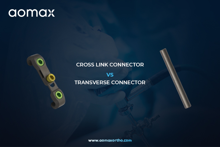
Cross Link Connector vs Transverse Connector - A Spine Implant Comparison
In posterior spinal fusion surgeries, cross-connectors are essential for linking bilateral rods, enhancing torsional stability, and reducing rod migration or parallel displacement. Two common options are the Cross Link Connector (Top Loading) and the Transverse Connector (Flat Rod). While both improve construct rigidity in thoracolumbar or cervical procedures, they differ in design, surgical application, and performance.
Design & Compatibility
The Top Loading Cross Link Connector features a pre-assembled or simple clamp design that loads directly onto standard round spinal rods (typically 5.5–6.0 mm) from above. It uses set screws for secure fixation and comes in adjustable lengths (e.g., 30–100 mm). Its titanium construction offers low profile and multi-axial adjustability in some systems.
In contrast, the Transverse Connector (Flat Rod) uses a flat connecting rod (often 4.0 mm) or interfaces with flat-profile elements. It connects rods or plates via side/front loading and is ideal for systems requiring rectangular bars or custom contouring. This design distributes stress differently and suits tighter anatomical spaces.
Surgical Ease & Stability
Top Loading excels in intraoperative efficiency: surgeons drop it onto rods after rod placement, minimizing tissue disruption and assembly time. It provides excellent anti-rotation strength with minimal ROM reduction in axial rotation.
Flat Rod Transverse offers superior contourability and a lower profile for complex deformities or revisions. It enhances rigidity in cervical constructs but may require more precise rod preparation.
Key Advantages
| Feature | Top Loading Cross Link | Flat Rod Transverse Connector |
|---|---|---|
| Loading | Top (fast, easy) | Side/Front (custom fit) |
| Rod Type | Round rods | Flat rods/bars |
| Best For | Standard fusions, speed | Deformities, low-profile needs |
| Stability Focus | Torsional (axial rotation) | Multi-plane + contouring |
Both reduce motion effectively, but clinical studies show the added benefit is often modest in single-level cases—choose based on patient anatomy and surgeon preference.
Conclusion: Select Top Loading for quick, reliable stabilization in routine cases. Opt for Flat Rod Transverse when contouring or profile matters most. Consult your implant system specs for optimal fusion outcomes. Always prioritize biomechanical needs for long-term success.
Get Connected:
+91 98989 50530 | ortho@advinhealthcare.com | www.advinortho.com
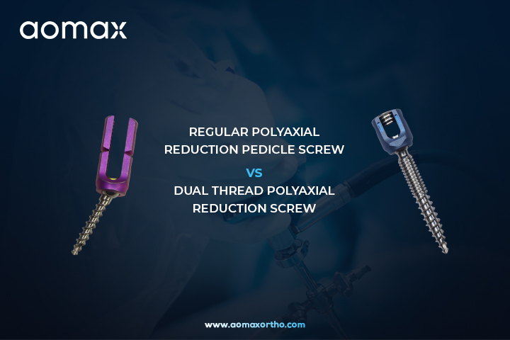
Regular Polyaxial Reduction Pedicle Screw vs Dual Thread Polyaxial Reduction Screw - Key Differences
In spinal deformity correction, spondylolisthesis reduction, and complex fusions, reduction pedicle screws play a critical role by allowing controlled approximation of the rod to the screw head. Two popular options are the Regular Polyaxial Reduction Pedicle Screw and the Dual Thread Polyaxial Reduction Pedicle Screw.
Regular Polyaxial Reduction Pedicle Screw features a standard single-thread design with a mobile polyaxial head (typically offering 25°–60° angulation). The extended reduction tabs or threaded mechanism enable powerful, controlled rod reduction. This design provides excellent versatility for rod capture in MIS or open procedures and simplifies alignment in challenging anatomy. However, in patients with variable bone density or osteoporosis, standard threads may offer comparatively lower initial purchase and pull-out resistance.
Dual Thread Polyaxial Reduction Pedicle Screw combines the same polyaxial reduction head with a dual-thread shaft — usually a coarse thread distally (for strong cancellous bone purchase in the vertebral body) and a finer thread proximally (for enhanced cortical engagement in the pedicle). This advanced thread pattern significantly improves insertion torque and pull-out strength (often 25–60% higher than single-thread designs in biomechanical studies). It also allows faster screw insertion due to the dual-lead effect while maintaining the reduction capability through extended tabs.
Quick Comparison:
- Rod Reduction & Alignment — Both excellent; polyaxial head offers similar multi-planar flexibility.
- Bone Purchase & Fixation Strength — Dual Thread superior, especially in osteoporotic or poor-quality bone.
- Insertion Speed — Dual Thread faster due to optimized thread design.
- Pull-out Resistance — Dual Thread provides higher biomechanical stability.
- Best Use — Regular for standard cases with good bone quality; Dual Thread for revision, osteoporosis, or when maximum anchorage is required.
Both screws are typically titanium for biocompatibility and strength. The choice ultimately depends on patient bone quality, pathology, and surgeon preference for construct stability.
In summary, while Regular Polyaxial Reduction Screws offer reliable performance and ease of use, Dual Thread versions deliver enhanced fixation and pull-out strength, making them a preferred upgrade in demanding spinal reconstructions.
Consult product IFU, surgical technique guides, and clinical evidence before use.
Get Connected:
+91 98989 50530 | ortho@advinhealthcare.com | www.advinortho.com

Revolutionizing Spinal Fusion - TLIF and PLIF Cages
In the fast-evolving field of spine surgery, interbody fusion cages have become essential tools for treating degenerative disc disease, spondylolisthesis, and spinal instability. These titanium or PEEK implants restore disc height, stabilize the lumbar spine, and promote solid bone fusion. Two of the most trusted options are the TLIF Cage and PLIF Cage – designed for precision, durability, and patient outcomes.
TLIF Cage (Transforaminal Lumbar Interbody Fusion)
The TLIF Cage is inserted through a single-sided posterior approach, minimizing muscle disruption and nerve retraction. Its bullet-nosed, lordotic design allows easy insertion even in collapsed disc spaces. Available in various heights (8–16 mm) and footprints, the cage features a large graft window and porous surface that accelerates osseointegration. Surgeons prefer TLIF for its minimally invasive compatibility, reduced blood loss, and lower risk of dural tears. Ideal for L4-L5 and L5-S1 levels, it delivers excellent segmental lordosis and long-term stability.
PLIF Cage (Posterior Lumbar Interbody Fusion)
The PLIF Cage is placed bilaterally from a direct posterior approach, offering maximum surface area contact with the vertebral endplates. Its rectangular or trapezoidal shape provides superior anterior column support and immediate rigidity. With a wide central canal for bone graft packing, PLIF cages are perfect for cases requiring robust fusion in patients with severe disc degeneration or revision surgeries. The design ensures parallel distraction and excellent radiographic visibility under fluoroscopy.
Why Choose These Cages?
Both implants are radiolucent (PEEK) or radiopaque (titanium) options with anti-migration teeth and FDA/CE-certified quality. They reduce recovery time, lower adjacent segment stress, and achieve fusion rates above 95%. Whether your case needs the unilateral precision of TLIF or the bilateral strength of PLIF, these cages deliver consistent, reproducible results.
At our facility, we combine cutting-edge engineering with surgeon feedback to make spine surgery safer and more effective. Ready to explore how TLIF and PLIF cages can transform your practice? Contact us today for product catalogs, surgical technique guides, or case studies.
Get Connected:
+91 98989 50530 | ortho@advinhealthcare.com | www.advinortho.com